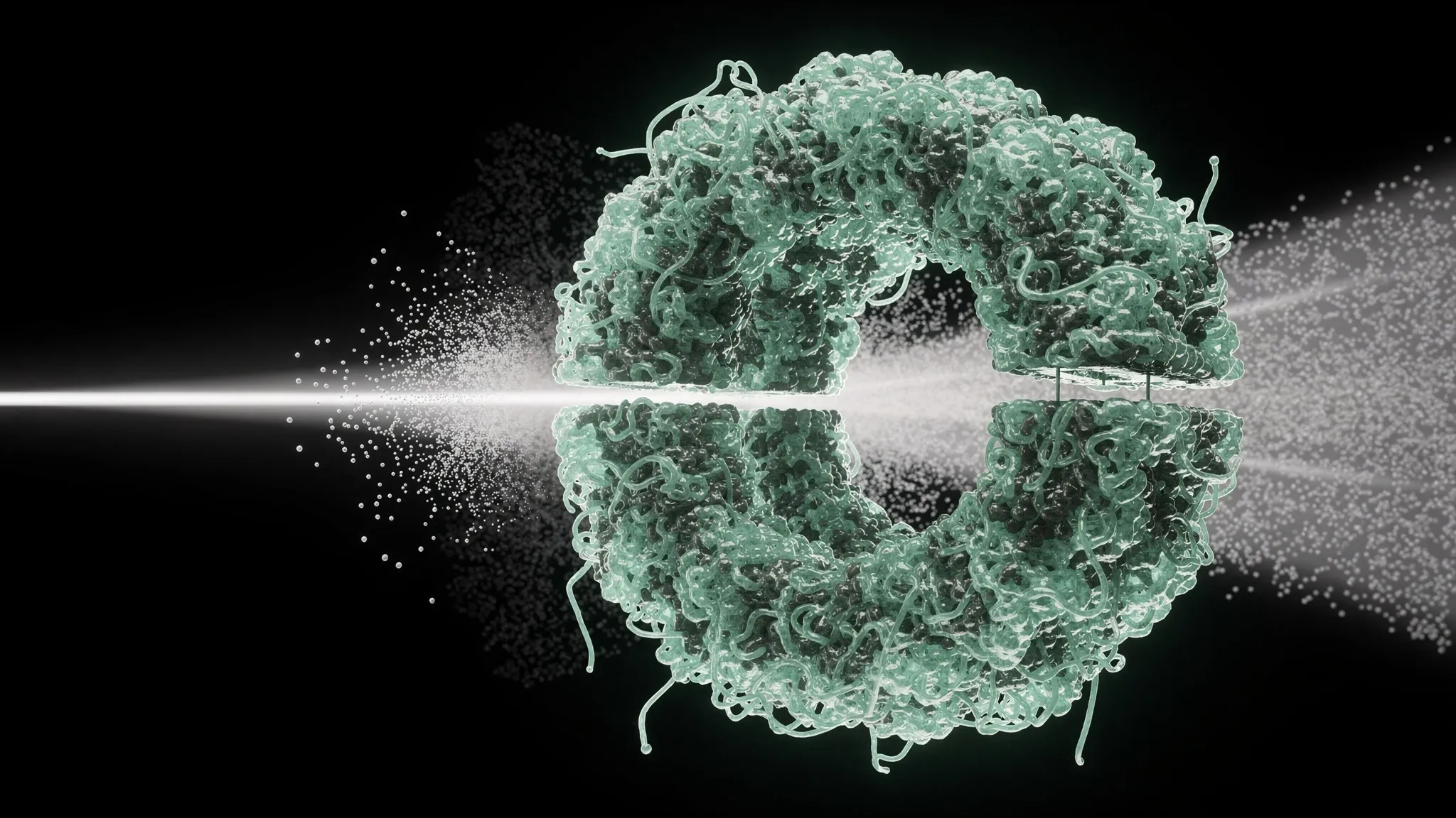
ONE PROTEIN, CAUGHT IN FLIGHT BY AN X-RAY FLASH
To see how a protein works, biologists would like to watch it change shape. Doing so means seeing a single molecule from many angles, with radiation of very short wavelength. The problem is that electrons and X-rays, the only probes strong enough, also damage the molecule they are imaging. Cryo-electron microscopy gets around this by freezing thousands of molecules in ice and averaging many noisy pictures.
X-rays dump about a thousand times more energy per scattering event than electrons. But there is a loophole, known as “diffraction before destruction”: with a pulse from an X-ray free-electron laser lasting only a few femtoseconds, the pattern of scattered light is recorded before the molecule has time to fly apart. The molecule needs no freezing and no support — it can be caught in flight. Until now, this had worked on large viruses. A single protein, imaged in 3D with hard X-rays, had remained out of reach.
Earthworm blood in a vacuum
A large international team led by Paul Lourdu Xavier and Henry Chapman at DESY in Hamburg, with the European XFEL and dozens of institutions worldwide, chose erythrocruorin: a giant haemoglobin, purified from the blood of about a hundred earthworms. It weighs 3.6 megadaltons and has a highly symmetric shape, two stacked rings with a hole in the middle.
The proteins were sprayed into fine droplets, neutralised and focused into a narrow beam travelling at about 100 metres per second through a vacuum chamber. Helium was added to cut the background glow from leftover gas. Across this beam fired pulses from the European XFEL in Schenefeld, Germany: X-ray photons of 6 kiloelectronvolts, squeezed onto a spot of 180 by 220 nanometres, each pulse lasting about 25 femtoseconds.
Each flash ionises about three quarters of the atoms in the molecule — but it is over before the resulting explosion has time to blur the picture.
270 million frames for one shape
- In 21 hours, the detector recorded 270 million frames.
- Only 188,557 of them — 0.07% — held a diffraction pattern, mostly from a single protein.
- The best 50,000 were sorted into 144 groups of similar viewing angles, which gave flat projection images at about 15 ångströms resolution.
- From 26,908 selected patterns, the team rebuilt the diffraction in three dimensions, then kept the 1,248 cleanest to compute a 3D map of the protein’s electrons at about 20 ångströms. It shows the central hole and the six-fold symmetry expected from the known structure.
A protein that shrinks in flight
All the 3D reconstructions came out more compact than the known crystal structure, especially along the short axis. The team traced this to the vacuum itself: out of water, the protein contracts. Computer simulations of the whole molecule agree — it shrinks by 2.5% in its first nanosecond, then more slowly. This variation between molecules is probably why the 3D image is blurrier than the 2D ones.
The authors list the next steps: protecting proteins from this collapse, with smaller droplets, a shorter trip or flash freezing, or by delivering them in an ultra-thin liquid sheet; and steadier, shorter, more powerful X-ray pulses. Simulations show that radiation damage will matter more as images get sharper.
Toward movies of single molecules
Twenty ångströms is far from seeing atoms. But the authors describe this as a goal pursued for almost three decades: a single, isolated protein, imaged in three dimensions with hard X-rays, without crystals or freezing. The promise beyond it is time — triggering a protein and catching it mid-motion, one flash at a time.