WATCHING THE BRAIN WORK THROUGH THE SKULL, WITH LIGHT AND SOUND
Functional MRI has transformed how we watch the human brain at work. But it senses oxygen-poor blood only indirectly, through its effect on water molecules, and it needs a bulky, costly, noisy, enclosed superconducting magnet. Other tools trade one strength for another: electrical recordings are fast but blurry in space; near-infrared light sees blood but scatters too much to give sharp images.
Photoacoustic tomography offers another route. Short laser pulses enter the tissue; when haemoglobin absorbs the light, it generates ultrasound waves, which sensors pick up to rebuild a 3D image. Oxygen-rich and oxygen-poor haemoglobin absorb different colours, so two lasers can tell them apart. The machine is more compact, cheaper and quieter than an MRI scanner, with no magnet at all.
The catch: in adults, functional photoacoustic imaging of the brain had only worked in people missing part of their skull after surgery. The bone dims both the light and the sound, distorts the waves, and creates echoes. And the signal changes caused by a mental or motor task are only a few per cent — easily drowned by a head that moves by a fraction of a millimetre.
A bowl of 3,072 ears
Yilin Luo, Rui Cao, Manxiu Cui, Lihong Wang and colleagues at the California Institute of Technology, with Danny Wang’s team at the University of Southern California, attacked both problems.
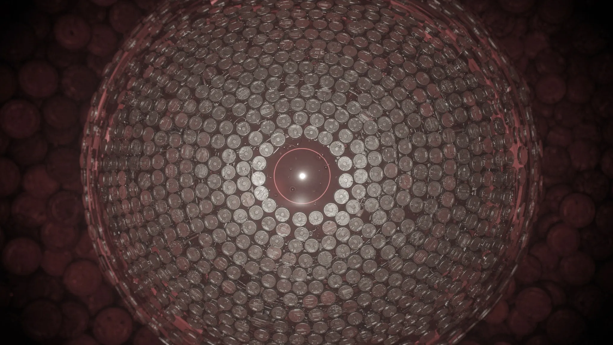
- They packed 3,072 ultrasound sensors into a hemispherical bowl around the head — the highest channel count to date — each with its own amplifier. They listen at a low frequency, 1 MHz, which crosses bone more easily.
- Compared with the team’s previous system, the estimated signal-to-noise gain through the skull is about 273-fold, enough to offset a skull that dims the signal by roughly a hundred times.
- Two lasers, at 1,064 and 755 nanometres, fire in turn. Each pulse yields a complete 3D image, 20 volumes per second, with no moving parts — so no blur from scanning.
- Each volunteer wore a personalised 3D-printed helmet, designed from a 3D scan of their head. Head motion typically stayed within about 0.1 millimetre and 0.1 degree over a 7.5-minute session.
On a human skull taken from a body and filled with gel, the system could still see a blood-filled tube placed inside, even though its signal was about 300 times weaker than outside.
Fingers, lips and a 7-tesla check
Four healthy men, aged 24 to 56, tapped the fingers of one hand or pursed their lips, 30 seconds on, 30 seconds off. Each also had a scan in a high-field, 7-tesla MRI as a reference.
- The method placed finger-tapping responses in the expected movement-and-touch area of the brain, on the side opposite the moving hand, matching the MRI.
- It told hand from mouth: in all 16 combinations of person, brain side and task, the photoacoustic map sat closer to the MRI map of the same task than to that of the other task.
- It repeated itself: in one volunteer, five sessions over two visits three months apart all landed in the same region.
- The two laser colours moved in opposite directions during the task, as expected from their different sensitivity to oxygen-rich and oxygen-poor blood. The light-and-sound signal at 1,064 nm tracked the MRI signal closely, with a correlation of 0.92.
The fine print
The photoacoustic maps sat on average 11.4 millimetres from the MRI ones, systematically shifted toward the brain’s surface. The authors list several possible causes they cannot yet separate: distortion by the skull, registration errors between machines, the brain shifting with posture, and session-to-session differences. Only four participants were tested, all men, on simple motor tasks, and the hair over the imaged area was shaved.
Lihong Wang declares a financial interest in three photoacoustic imaging companies, which the authors state did not support this work.
The team sees a platform that could sit next to other tools — electrical recordings, brain stimulation, brain–computer interfaces — that are hard to combine with an MRI magnet. With more compact lasers, they hope to study the brain in more natural settings.