BARE NERVE FIBRES SKEW THE MRI READING OF MYELIN LOSS
In the brain’s white matter, many nerve fibres (axons) are wrapped in myelin, a fatty sheath. When myelin is destroyed, as in demyelinating diseases, doctors and researchers follow the damage with quantitative MRI: measures of how water diffuses, how fast the signal fades, or how myelin exchanges magnetization. These measures are usually read as if every axon in a voxel — one image cell — were sheathed. Fibres without myelin are assumed to count for little.
Ali Abdollahzadeh, Els Fieremans, Dmitry Novikov and colleagues, at New York University, the University of Eastern Finland and Weill Cornell Medicine, checked that assumption.
Mice, MRI and a microscope
The team reanalysed data from 36 female mice. Twelve ate normal food; the others ate food laced with cuprizone, a compound that destroys myelin, for 6 or 12 weeks, followed by 6 weeks of recovery. Their brains were scanned by MRI at 0, 6, 12 and 18 weeks, and slices of the corpus callosum, the white-matter region where cuprizone strips the most myelin, were imaged with an electron microscope at a resolution of 2.36 nanometres.
Bare axons are hard to outline automatically: they are thin and surrounded by a single faint membrane. The team trained a neural network that started from one hand-labelled image and was corrected step by step, until it could sort every image into myelin, sheathed axons, bare axons and the space between them.
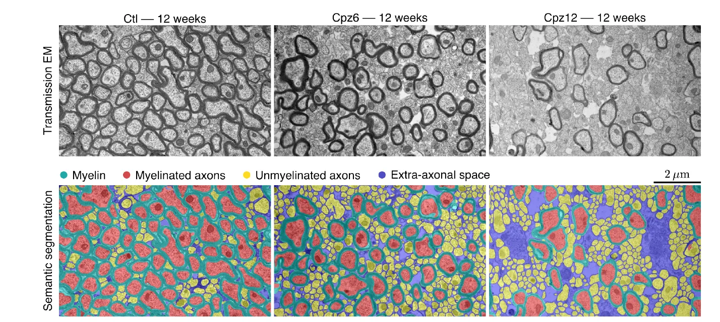
Corpus callosum after 12 weeks in a control mouse (left) and in mice fed cuprizone for 6 or 12 weeks. Top: electron microscopy; bottom: automatic segmentation into myelin (teal), myelinated axons (red), unmyelinated axons (yellow) and extra-axonal space (blue). Scale bar: 2 µm. — Figure 3 (top rows), Abdollahzadeh et al. (2026), arXiv:2611.24487, CC BY 4.0.
Fewer sheaths, not fewer fibres
Under cuprizone, the amount of myelin and the share of sheathed axons fell, while bare axons and the space between fibres grew. The total volume of axons stayed roughly constant, and their diameters did not change: sheathed axons averaged 0.55 µm across, bare ones 0.23 µm. The fibres seem to have survived; what changed was the share that kept their sheath. Myelin partly returned after cuprizone was stopped.
What the MRI was really measuring
Two results change how the scans should be read.
- The axonal water fraction, a diffusion-MRI measure, is usually read as the space inside myelinated fibres. Compared with the microscope, it lines up with all axons: a straight line through the points should start from zero if MRI and microscope measure the same thing. Against the total axon volume, it nearly does (−0.04); against sheathed axons alone, it starts well above zero (0.20). At the short diffusion time used here (16 milliseconds), water inside bare fibres counts too.
- The g-ratio, the inner radius of a fibre divided by its outer radius including myelin, measured for sheathed axons alone, did not change in the treated mice: the surviving sheathed fibres kept their proportions. The g-ratio estimated by MRI tracked the microscope well (correlation 0.77) only when bare axons were counted, as fibres with a g-ratio of 1, since without a sheath the inner and outer radius are the same. A rising MRI g-ratio thus reflects a growing share of bare fibres, not thinner sheaths on each fibre.
The authors also show mathematically that mixing different fibres in one voxel pulls the MRI-style g-ratio down, by up to 8% in tissue rich in bare axons. Their correction for this effect overshot in practice, so variability alone does not explain all the gap between MRI and microscope.
A matter of timing
The water inside bare axons counts only because the measurement is fast. Over longer diffusion times, typical of clinical scanners, that water exchanges with the space around the fibres, and the bare axons’ share fades: clinical scans may therefore be less sensitive to them. The authors argue that telling sheathed from bare fibres matters most when myelin is lost while axons survive, as in the early demyelinating or remyelinating stages of multiple sclerosis. This work, though, is in mice.