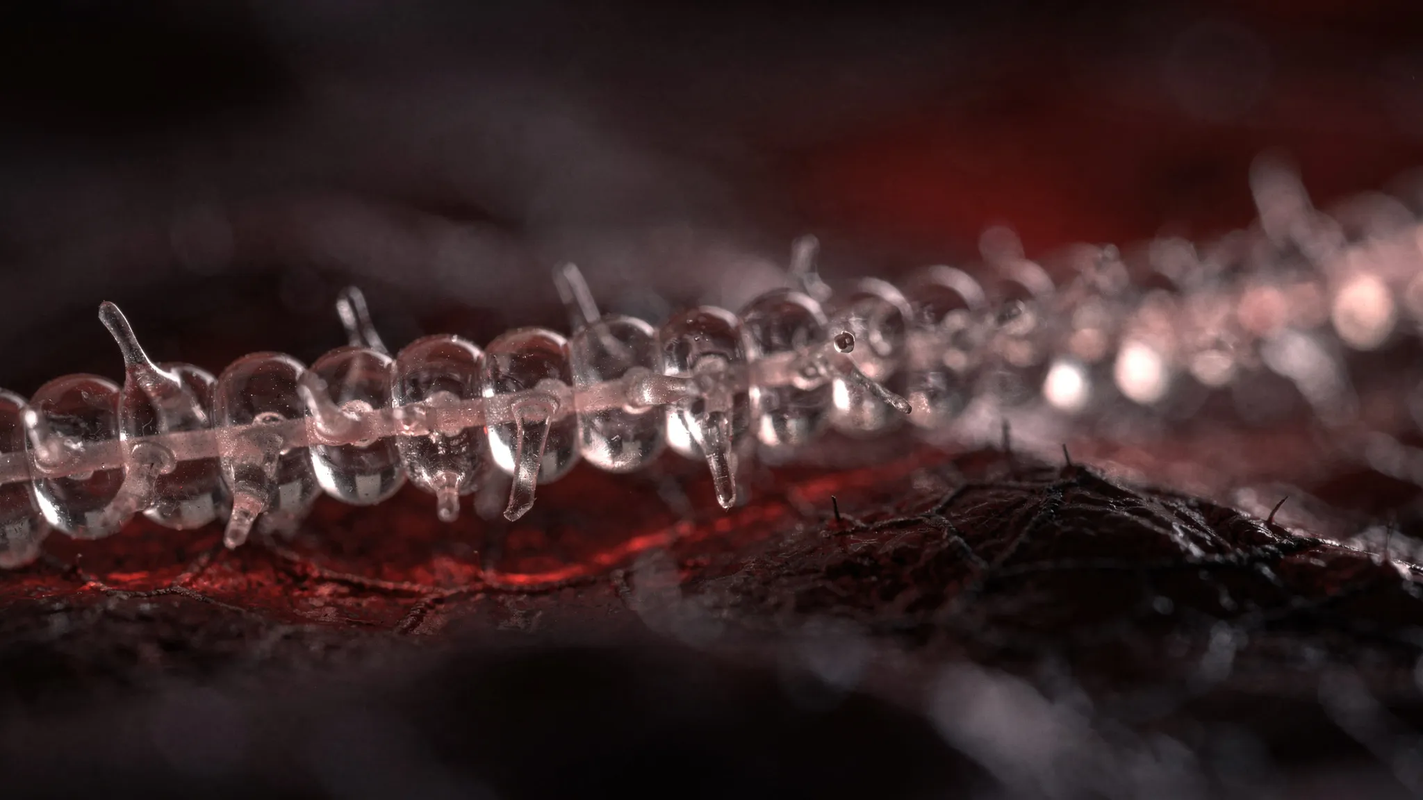
THE BODY'S HIDDEN PUMPS, SIMULATED FROM HEAD TO TOE
Alongside the blood vessels runs a second, quieter network: the lymphatic system. Every day it returns 2 to 3 litres of fluid that has leaked out of the blood into the tissues, together with proteins, fats and immune cells, back into the veins. Most of it ends in the thoracic duct, which empties into the junction of two large veins at the base of the neck. The same network also drains the central nervous system.
Lymph has no heart. It is pushed forward by lymphangions: short segments of vessel wrapped in muscle and closed by valves at each end, which contract like miniature pumps. When this pumping fails, fluid builds up — a condition called lymphoedema — and it is implicated in immune and metabolic diseases too.
From one vessel to the whole body
Existing models describe one lymphangion, a short chain of them, or a small regional network, often in mice. The only whole-body model of the human lymphatic system was passive and did not compute pressures. Chia-Feng Chang, Sabee Molloi and colleagues in the Department of Radiological Sciences at the University of California, Irvine, set out to fill the gap in two steps, published as two companion papers.
The skeleton. In the first paper (arXiv:2610.12750), they took an anatomical map of human lymph vessels — 1,010 junctions and 1,126 vessels, reviewed by a clinical anatomist — and cut it into 3-millimetre lymphangions and then into about 89,000 segments. Vessel sizes were set by a classic rule linking flow and radius, Murray’s law. With the muscles switched off, the radii and pressures that came out matched anatomical and clinical ranges without being fitted, and about 49% of the lymph load was reabsorbed or held back by lymph nodes before reaching the veins.
The pumps. In the second paper, the one discussed here, they switched the muscles on. Each segment now contracts on a rhythm specific to its body region, its wall relaxes with a realistic delay, its valves open and close with the local pressure — and the exit valve into the veins opens on each in-breath and shuts on each out-breath.
A pump that finds its own rhythm
The team did not fit the shape or the strength of the contractions to any data. Starting from rest, the network first filled up for 50 to 100 seconds, then settled by itself into a steady pumping cycle, beating in time with the breath at the junction with the veins:
- pressure at the outlet of 7.5 to 12.3 mmHg;
- drainage of 35 to 50 cubic millimetres per second;
- both within published clinical ranges, and both rising with the strength of contraction.
Two rhythms can be told apart. At the junction with the veins, breathing dominates, with a swing of 1 to 2.5 mmHg. Inside the thoracic duct, the contraction dominates, with a swing of about 1 mmHg. That helps interpret clinical catheter readings: what they record depends on where the catheter sits.
What limits the pump
A lymphangion sealed at both ends could raise its pressure by about 11 mmHg. In the network, it achieves only about 14% of that ceiling: as the muscle squeezes, the pressure in the vessel and the stiffness of its wall push back, and the radius changes by only a few percent per beat. In the authors’ words, the pump is limited “more by its load than by its drive”.
A second conclusion holds whatever the unknown parameters: no single contraction strength fits every vessel. The pumps will have to be tuned vessel by vessel — the subject of a planned follow-up study.
The route from the brain
The authors also looked at the neck, through which fluid draining from the brain reaches the veins. When they raised the pressure in the veins from 3 to 15 mmHg, the pressure in the deep cervical lymph node rose from 8.6 to 21.1 mmHg with weak contractions — passing on slightly more than the full rise. With strong contractions, only 78% of the rise came through. Contraction, in other words, buffers drainage from the neck against venous pressure. The authors link this to brain swelling after head injury, where — in mice — both venous pressure rises and lymphatic contraction weakens.
The model also computes the pressure at the base of the neck that models of brain-fluid drainage currently have to assume.
A foundation, not yet a clinical tool
The authors are clear about the limits. Human measurements of the muscle, wall and valve properties do not yet exist across the body, so the pumping side relies on mouse and rat data rescaled to human size, and the exit resistance on pig measurements. Contraction strength is the same everywhere, the valves are evenly spaced, the deep neck region is a single point of the map, and none of the clinical statements has been tested against patient data. They describe this healthy, whole-body model as a foundation on which disease states can later be studied.