MUONS SEE INSIDE A CHEST FOR THE FIRST TIME
Muons are the electron’s heavier cousins, about 200 times more massive. That weight lets fast muons cross great thicknesses of matter while losing little energy. Cosmic rays rain them down on Earth’s surface all the time, and physicists have turned them into an imaging tool: muography. It has probed volcanoes, glaciers and archaeological sites, and famously revealed a large unknown void inside the Great Pyramid of Giza.
A second technique, muon scattering tomography, measures how much each muon is deflected as it crosses an object. Dense, heavy materials deflect muons strongly, which is why this method is used for cargo inspection and the search for shielded nuclear material.
The faintest possible signal
The human body is the opposite case. Soft tissues are made of light elements and have similar densities. The deflections they produce are orders of magnitude smaller than those of uranium or steel. Whether muon tomography could see anything in soft tissue was an open question.
The appeal is real: muons of a few billion electronvolts pass through the body depositing little energy. The authors do not see them replacing X-ray scanners, but as a possible complement where low dose or repeated imaging matters. The catch is the source: the natural cosmic flux is far too weak, and intense muon beams exist only at large accelerators.
A plastic chest in a CERN beam
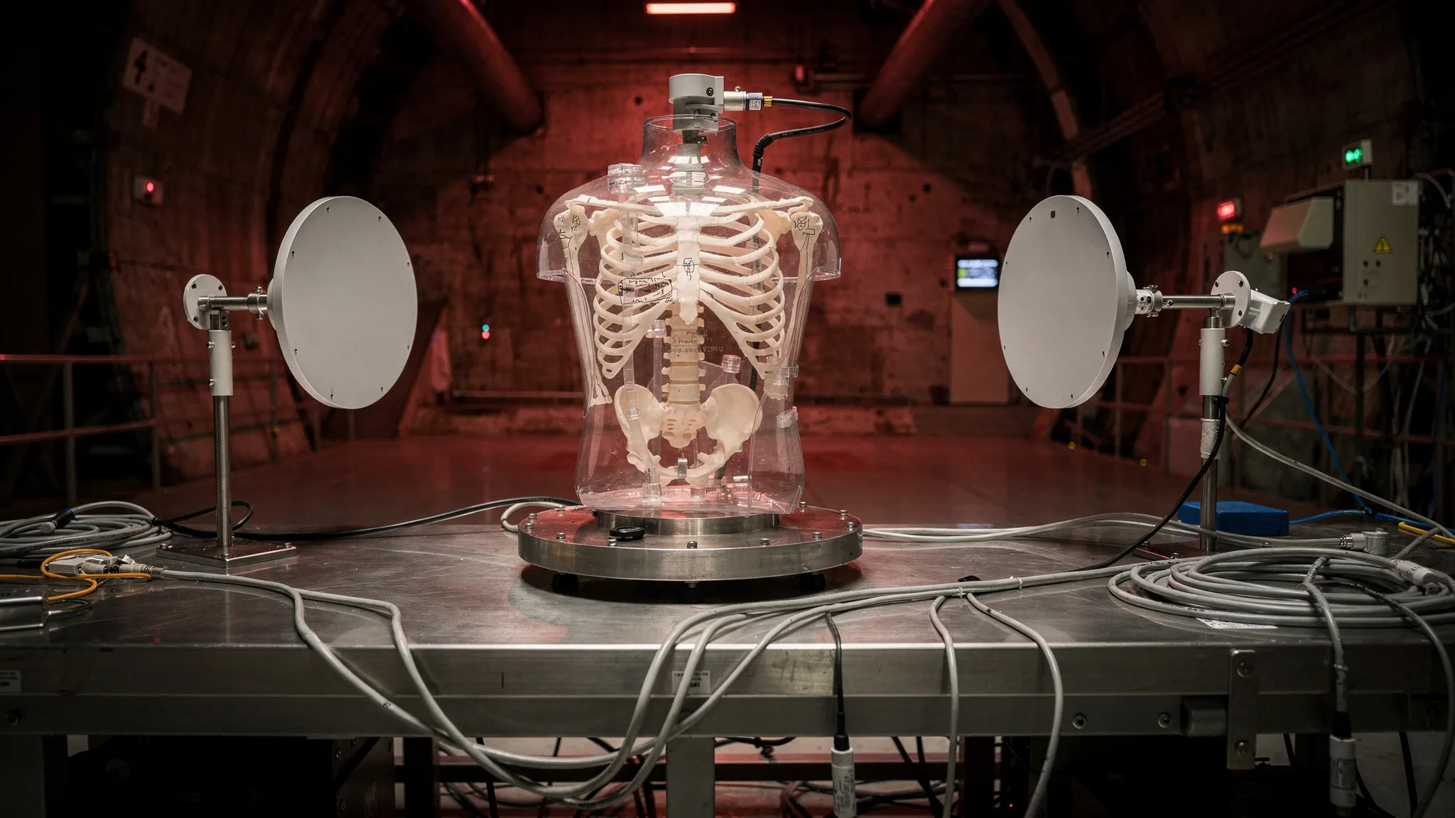
Dipanwita Banerjee of CERN and colleagues from ETH Zurich and the Institute of Corpuscular Physics in Valencia borrowed an Alderson thorax phantom from Geneva University Hospitals. It is a dummy used in clinical dosimetry: a tissue-like plastic body with lungs, a heart, a liver and a full skeleton made of bone-equivalent material.
In April 2026, they placed it in a 2 GeV/c muon beam at CERN’s Proton Synchrotron. Two detectors in front and two behind recorded the path of every muon in and out, giving its deflection. They rotated the phantom by hand to seven angles and kept 293,035 muons.
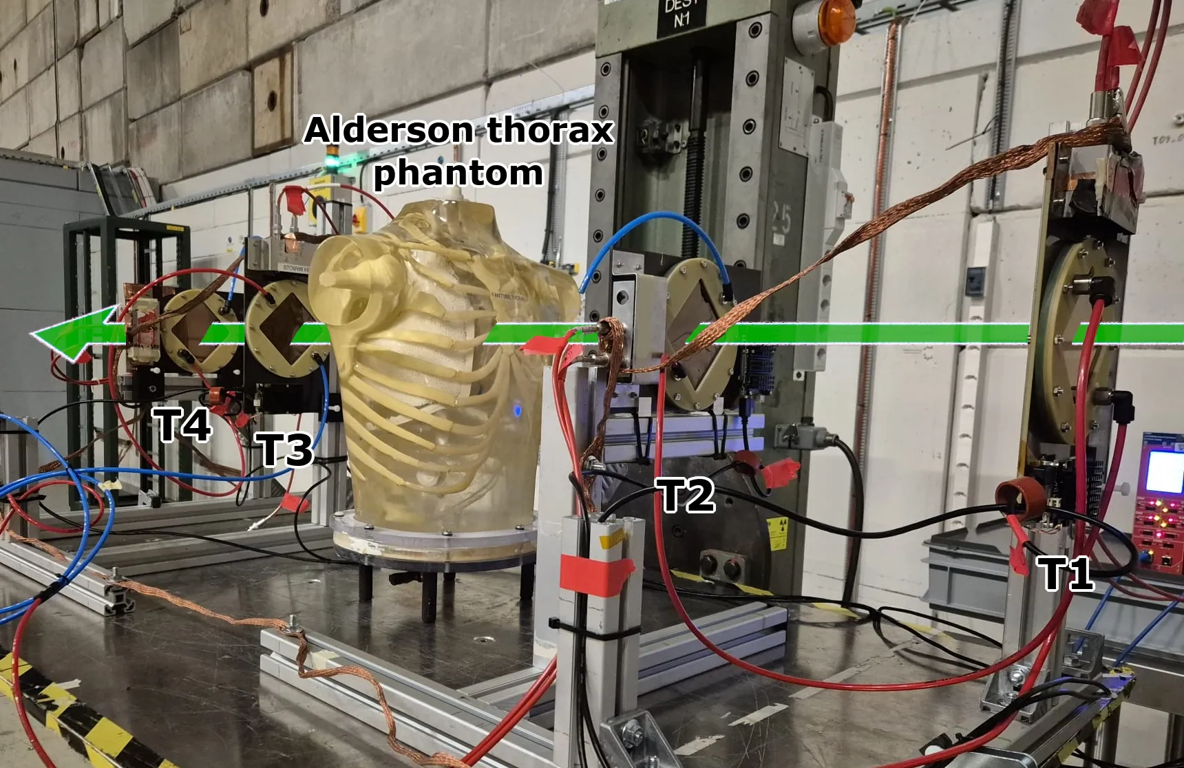
The Alderson thorax phantom between the upstream (T1, T2) and downstream (T3, T4) trackers on CERN’s T9 beamline. — Figure 1, Banerjee, Banto Oberhauser, Crivelli & Molina Bueno (2026), arXiv:2610.16647.
The raw reconstruction is very noisy. So the team added a 3D neural network that removes noise, trained only on 50 simulated phantoms with randomly resized organs — never on the real data.
Within 1% of the truth
The beam was narrow, about 6 by 4 centimetres. Only a small region was seen from all seven angles: a block of about 8 centimetres around the heart, 0.8% of the body. All the precise results apply there.
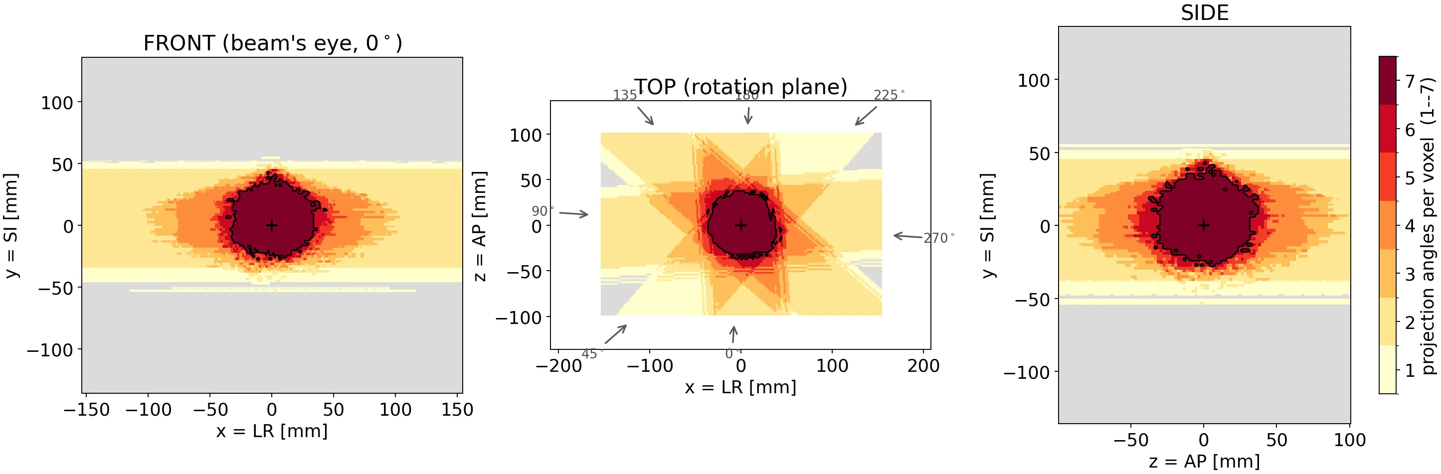
Which parts of the phantom the beam actually reached: the dark red core was seen from all seven angles, about 0.8% of the body. — Figure 2, Banerjee, Banto Oberhauser, Crivelli & Molina Bueno (2026), arXiv:2610.16647.
- In that core, the measured scattering density was 1.304 × 10⁻⁴ rad²/m, against about 1.297 × 10⁻⁴ expected from simulation: agreement better than 1%, with no recalibration.
- Changing how strictly stray tracks were filtered shifted the raw image by 15%, but the network’s output by less than 1%.
- Outside the core, ribs, sternum and spine showed up roughly where they should, with close to the right density for bone, but stretched into streaks.
Equally telling is what the method missed. The phantom’s heart wall contains small air-filled defects of 9 to 14 millilitres. They were not recovered: their signal, about 10%, sits at the statistical noise floor. The authors estimate that ten times more muons would put them at the edge of detection.
Two microgray
Each voxel of the core was crossed by about 58 muons. The authors estimate the local dose at about 2 microgray, and about 20 with ten times more muons. For scale, they cite about 0.1 millisievert for a chest X-ray and about 6 for a chest CT scan — while warning that these quantities are not directly comparable and that a clinical assessment would need a full dose calculation.
From the accelerator to the clinic?
According to the authors, this is the first experimental demonstration of muon scattering tomography on soft tissue. A clinical system would need many more angles and alignment precise to below a millimetre. Its natural targets would be lesions with a clear density contrast, such as a solid nodule inside air-filled lung, or calcified tissue. Everything now depends on the muon source: the team points to compact, laser-driven accelerators, which promise beams from room-sized facilities.