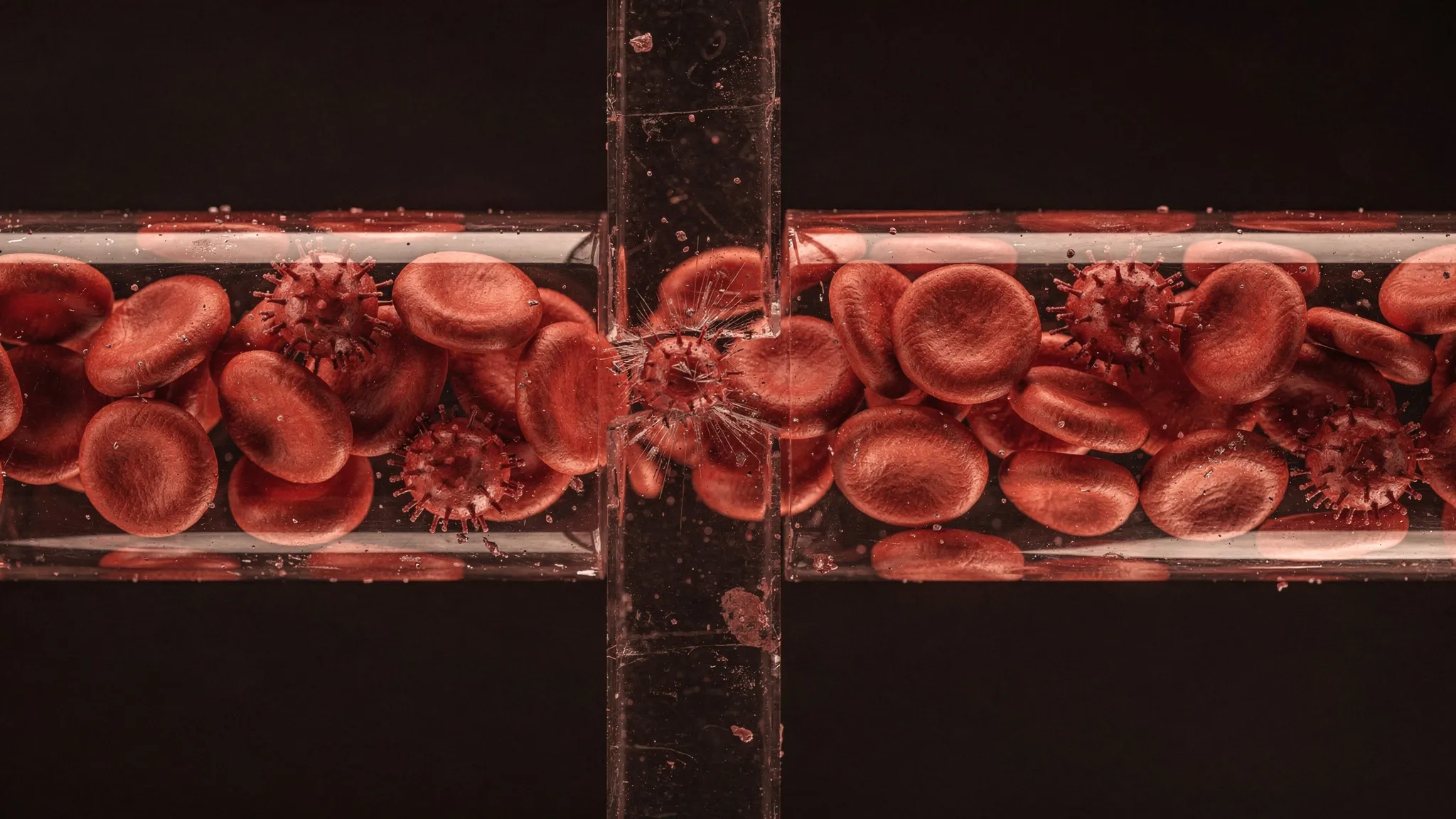
SPIKY RED CELLS GET STUCK IN A VIRTUAL SPLEEN
Red blood cells kept for transfusion do not stay smooth discs. They turn into echinocytes: first discs with an irregular rim (stage I), then discs with a scalloped edge (stage II), then compact cells covered in small spikes (stage III). Later they shed little vesicles of membrane and end up as small, rounder cells. The spikes come first; the membrane loss follows.
Stage III cells and these small, rounder cells form a group whose projected area is 15 to 20% below that of the others. Its share of stored blood rises from about 5% after 3 days to about a quarter after 42 days. When stored blood was pumped through isolated human spleens, 61% of these cells were cleared within 70 minutes, against 15% of all the stored cells. In 31 healthy volunteers, units richer in them tended to leave fewer transfused cells in circulation a day later.
Squeezing through a slit
The spleen filters blood through tiny slits in the walls of its veins, about 1.9 micrometres long, with openings of 0.25 to 1.2 micrometres. Red cells that can no longer squeeze through are held back and can then be swallowed by macrophages, immune cells that clear debris.
Earlier models tied passage to just two numbers: the cell’s membrane area and its volume. To straddle the wall, a cell must split its volume into two lobes, one on each side, and that takes enough membrane. But in real stored cells, spikes and membrane loss arrive together. Zhaojie Chai, Jianlu Zheng, He Li, Ming Dao and George Em Karniadakis, at Brown University, MIT and the University of Georgia, asked whether the spikes alone could hold a cell.
Building a spiky cell on a computer
The team used a model in which the cell has two layers: an outer lipid membrane and an inner skeleton, each a mesh of 2,562 points, tied together by short links. In a normal disc, each membrane point is linked to its partner on the skeleton. To make an echinocyte, only 462 pairs stay linked, along lines that trace the edges of an icosahedron (a solid with 20 triangular faces), which leaves 20 free patches of membrane, and the skeleton shrinks. Each patch then lifts into a spike, on average 1.28 micrometres high in stage III.
Crucially, every cell keeps exactly the same membrane area and volume as the disc. And the shrinking skeleton already stretches the links before the cell meets anything: their typical length rises from 0.53 of their maximum in the disc to 0.81 in stage III.
Stuck at 37 to 50 times the pressure
Each model cell was pushed into a slit 4 micrometres wide and 1.2 micrometres high, with the pressure raised step by step. The disc passed at 1.5 or 2 pascals, a stage I cell at 4 and a stage II cell at 12.5 or 15. The stage III cell was still held at 75 pascals in every run, the highest step before the simulations broke down: 37 to 50 times the pressure that let the disc through. With the disc’s softer skeleton, it was still held at 50 pascals, so its shape, more than its stiffness, does the holding.
The comparison with smooth cells is the clearest. A smooth cell gets stuck once it has lost between 5 and 7.5% of its membrane, when its area no longer suffices for two lobes. A spiky cell that had lost 5% of its membrane, whose volume was no harder to split into two lobes with its membrane than that of a smooth cell that slipped through at 1.5 or 2 pascals, was held up to 75 or 100 pascals.
The links that hold
What holds a spiky cell is its links. In held spiky cells, 53 to 77% of the links between membrane and skeleton were stretched beyond 90% of their maximum length, against at most 2.4% in cells that passed at the next step. Smooth cells held for lack of membrane kept their links slack.
The team then ran a direct test. Starting from a stage III cell stuck at 50 pascals, they lengthened only the links, changing nothing else. Left as they were, the cell stayed put. At 1.5 times their length, it crept forward by a fraction of a micrometre. At twice their length, it passed in six runs out of eight; at 2.3 to 2.4 times, in every run.
Why filters missed it
Older experiments had found that stage II and III echinocytes pass through 2.6-micrometre filter pores like normal cells. The model explains this: in a 3.2-micrometre pore, all stages pass at pressures well below those reached across such filters. Even in channels 5 micrometres wide, like those of the team’s microfluidic device (a chip carved with tiny channels), stage III cells entered at 8 to 16 times the disc’s pressure — a far weaker sorting than the slit’s.
Applied to the mix of shapes in real stored blood, the model slit holds, above 15 pascals, only this group of small cells, stage III included: 0.25% of the modelled cells on day 3, 18.6% on day 42. Perfused spleens, too, cleared these small cells far more than stored cells as a whole (61% against 15%). The authors compare only this ordering, not the percentages, because the pressures in their model are not calibrated against a real spleen.
The study also has a practical twist. Treatments that “rejuvenate” stored cells restore their shape. In the model, removing the spikes helps a cell through only while its volume still fits its remaining membrane — a prediction that can be tested on stored cells sorted by shape, in slits narrower than a micrometre.