SKULL IMPLANTS THAT LOOK RIGHT BUT CANNOT BE FITTED
After a decompressive craniectomy — an operation that removes part of the skull to relieve a swelling brain —, a head injury or the removal of a tumour that takes bone with it, a patient is left with an opening in the skull. It is closed with an implant shaped to that patient’s anatomy, which must protect the brain and restore the skull’s shape. Whether it fits is settled in the operating room.
These implants are still drawn by hand by specialists in computer-aided design. Each case is slow and expensive, which limits how many patients can get one. Networks trained specifically for this task do well on public benchmarks, but they need training data for it. Foundation models — large models pre-trained on huge collections of 3D shapes — promise to skip that step: a skull with a hole is just a shape to complete.
Judged like a surgeon would
Marek Wodzinski and colleagues, at AGH University of Krakow and the Sano Centre for Computational Medicine in Poland, audited three publicly released models, used as they come:
- MSN-FM, which describes itself as the first multimodal foundation model for completing medical 3D point clouds — a shape handed over as a scatter of points, with no surface and no inside — pre-trained on 100,000 medical shapes and able to take text;
- MGPC, trained on a million pairs, with text and image inputs;
- AdaPoinTr, trained on 55 categories of everyday objects — none of them a skull — as a non-medical control.
They used 239 skulls: 210 with holes carved artificially from complete skull scans, and 29 real post-craniectomy cases with the implant an expert actually designed. Before running a single model, they fixed two criteria:
- Beat doing nothing: compare each output with simply handing back the patient’s remaining bone. The removed bone is a thin shell next to the bone that stays, and the score counts the missing region as covered wherever material lies within 2 mm of it, so simply sitting next to the hole’s edges earns points for free.
- Physically fit: the predicted material must sit in the hole without cutting into the remaining bone (1-millimetre tolerance) and cover at least as much as doing nothing.
As a yardstick, they built a model trained for this exact task, picked among 138 variants for its score on these very 210 cases, which flatters it — a best-case ceiling rather than a fair competitor — and trained only on artificial holes, unlike the 29 real skulls — and spent the second half of the study trying to break their own conclusion.
Zero out of 239
- On the 210 artificial holes, none did better than doing nothing. Measured as the share of the missing region recovered to within 2 mm, their median advantage over the remaining bone was negative: AdaPoinTr −0.036 (3.6% of the missing region less than the bone alone), MGPC −0.130, MSN-FM −0.156. The task-specific model gained +0.745.
- Medical pre-training did not help: MSN-FM came last, behind a model trained on everyday objects.
- On the 29 real cases, MGPC did beat doing nothing, on 24 of 29 — the only result beyond chance. The authors say that a model which loses on artificial holes but wins on 29 real skulls does not settle the clinical question.
- Implantable outputs: 0 of 239 for each of the three models, against 238 of 239 for the task-specific model. Across 2,868 combinations of model, setting and skull, none stayed clear of the bone at 1 mm. The models’ material ran a median of 5.4 to 6.6 mm into the patient’s own bone, against 0.55 mm for the task-specific model.
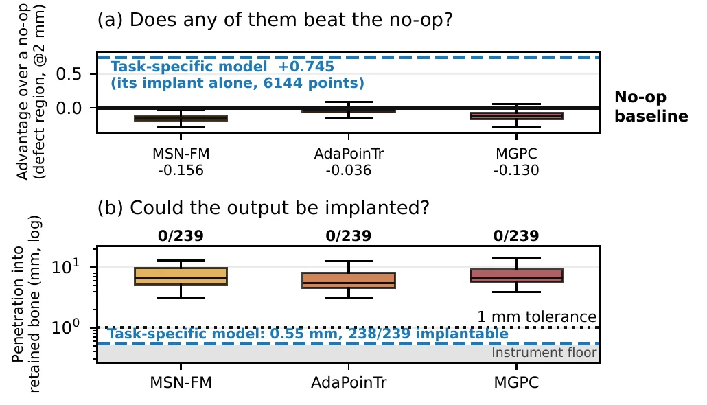
(a) Gain over doing nothing: the three models sit below zero, the task-specific model far above. (b) Depth of penetration into the remaining bone, with 0/239 implantable for each. — Figure 1, Wodzinski et al. (2026), arXiv:2610.30035, CC BY-SA 4.0.
The collisions are in the shape of the outputs, not in their placement: moving them changed almost nothing.
Pretty is not placeable
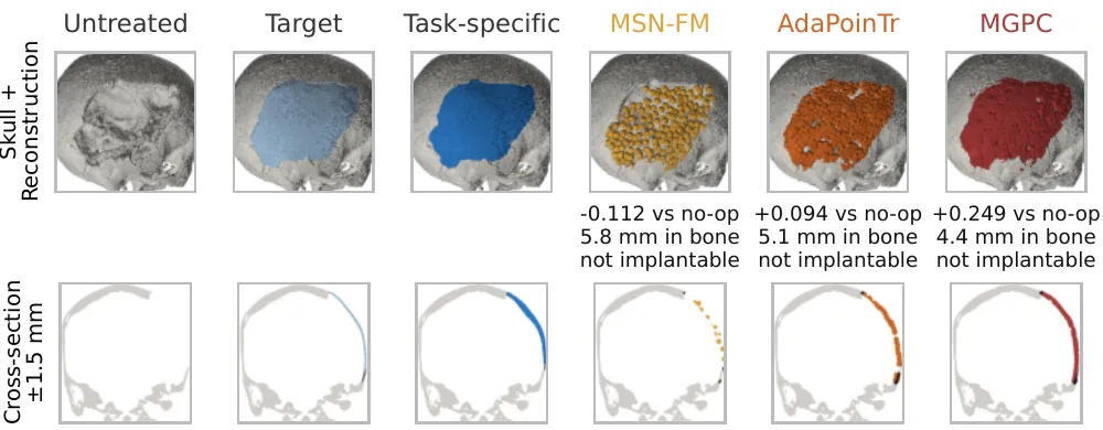
One real case seen from outside and in cross-section. The three models cover the hole, but all run into bone the patient still has. — Figure 2, Wodzinski et al. (2026), arXiv:2610.30035, CC BY-SA 4.0.
AdaPoinTr returns a clean surface that a reader would accept as a skull, yet it is no more placeable than the others. And thickness is lost from the start: with 6,144 points spread over a skull, neighbouring points are 2.56 mm apart on average, so the two faces of a 3-mm plate cannot be told apart. That is a property of the point-cloud format, not of any single model. Real implants are thin: 11 of the 29 were under 3 mm.
The text inputs that these models advertise turned out to be decorative. In MSN-FM, 97.5% of the text signal is exactly zero.
A trust problem
“Look at the pictures and the models have done their job: the defect is covered, the head has its shape back,” the authors write. Measure the same outputs and none can be put in a patient. A diffuse cloud warns you; a clean cap that scores unremarkably and still cuts into bone does not. Fixing these outputs by hand, skull by skull, is the manual work the models were meant to remove.
Their conclusion: as released, the shape-completion foundation models they could obtain “cannot be trusted” and “are not usable in clinical practice”. The study did not test fine-tuning on skulls, no clinician reviewed the outputs, and 29 real cases cannot settle a clinical question on their own. The paper has been accepted at a workshop of the ACCV 2026 conference.