A TUG-OF-WAR THAT TELLS A FLY EGG WHERE ITS BACK IS
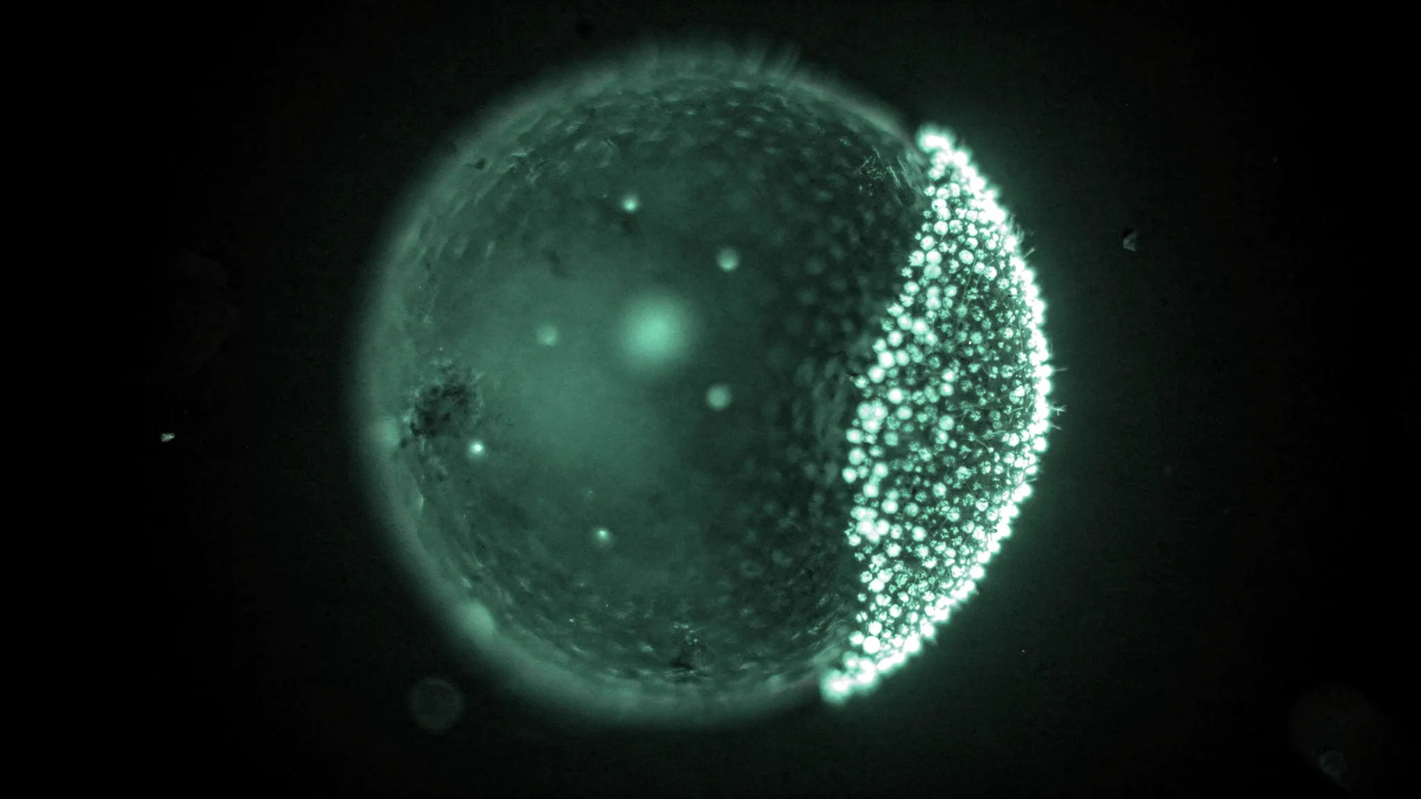
Long before it becomes an embryo, the egg cell of a fruit fly — its oocyte — has to decide which end is the front and which is the back. Part of this job falls to a messenger RNA called oskar. Made in the neighbouring “nurse” cells at the front, it is shipped into the oocyte, where it teams up with a protein called Staufen to form tiny particles. Early on, these particles are scattered throughout the cell; a few hours later, most of them form a cap at the back pole, the spot where the future germ cells will be specified.
How do they get there? Mohammadhossein Firouznia and Michael Shelley at the Flatiron Institute in New York, with Vladimir Gelfand’s team at Northwestern University in Chicago, combined live microscopy, genetics and a physical model to find out.
A tug-of-war at the edge
Earlier work by the same groups had shown that two motor proteins compete for oskar at the edge of the cell, its cortex. Myosin-V walks on actin filaments and holds the particles at the cortex. Kinesin-1 walks on microtubules and pulls them back into the cell. Actin is the same everywhere along the edge. Microtubules are not: as the oocyte grows from about 27 to 77 micrometres long in roughly 12 hours, they become much sparser toward the back.
So the release of oskar should be strong at the front and weak at the back. The team measured it directly. They fused Staufen to a protein that switches from green to red under violet light, flashed three spots of the cortex — front, side and back — and watched the red glow fade.
- Particles land on the cortex at about 2.6 times per minute.
- They leave at about 1.15 per minute at the front, 0.80 on the side and only 0.24 per minute at the back.
- At the back, capture is therefore about 20 times faster than release.
A droplet waiting to happen
There is a complication. Oskar particles tend to stick together, like oil in water: above a certain concentration, they separate into a droplet. The model captures this with a classic formula for mixtures, and the fitted stickiness turns out close to that of the “P granules” of C. elegans, droplets measured in earlier work.
The outcome depends on just two numbers: the total amount of oskar in the cell, and the ratio of release to capture at the cortex. With the measured values, the model reproduces the wild-type egg: a cap at the back and a uniform interior. It also predicts what happens when the balance is broken, and the experiments followed suit:

Where oskar ends up, from left to right: normal oocyte (cap at the back), kinesin-1 knocked down (whole edge), hyperactive kinesin-1 (cytoplasmic clump) and extra Patronin (droplet). Scale bars, 10 µm. — Figure 4D, Firouznia, Lu, Lakonishok, Farhadifar, Gelfand & Shelley (2026), arXiv:2610.10916, CC BY 4.0.
- No kinesin-1, so no release: oskar coats the whole edge, with no preference for the back.
- Hyperactive kinesin-1, a mutant that never switches off: oskar is pulled off the cortex and condenses into a droplet in the cytoplasm.
- Extra Patronin, a protein that stabilises microtubules and so strengthens kinesin’s tracks: again, a droplet.
Robust, but only up to a dose
At the natural amount of oskar, the release-to-capture ratio can vary about eightfold without the RNA falling into a droplet. But doubling the amount of oskar is enough to trigger the droplet. The authors suggest that the quantity of oskar a fly makes may itself be tuned by evolution — enough to build the cap, not so much that the excess competes with it.
In the mutant eggs, the droplet also stayed close to the back pole from one stage to the next, which the team explains by the oocyte growing mainly at its front. Later in development, strong swirling flows appear in the egg; whether they help repair a misplaced cap is the next question on the authors’ list.