A PICOSECOND PULSE THAT KILLS FEWER CELLS
More than half of all cancer patients worldwide receive radiotherapy at some point in their treatment. Its main limitation is the harm it does to healthy tissue around the tumour. One promising answer is FLASH radiotherapy: delivering high doses in a much shorter time, at mean dose rates of tens to hundreds of grays per second (the gray is the unit of radiation dose absorbed by tissue). It reduces damage to normal tissue while controlling tumours as well as conventional treatment. But what exactly triggers this effect, and why, is still debated — transient oxygen depletion, recombination of free radicals and mitochondrial responses have all been proposed. In cell cultures, the effect has proved elusive.
A team from Queen’s University Belfast and the UK’s National Physical Laboratory took the idea to an extreme: deliver the whole dose essentially at once.
Electrons from a laser
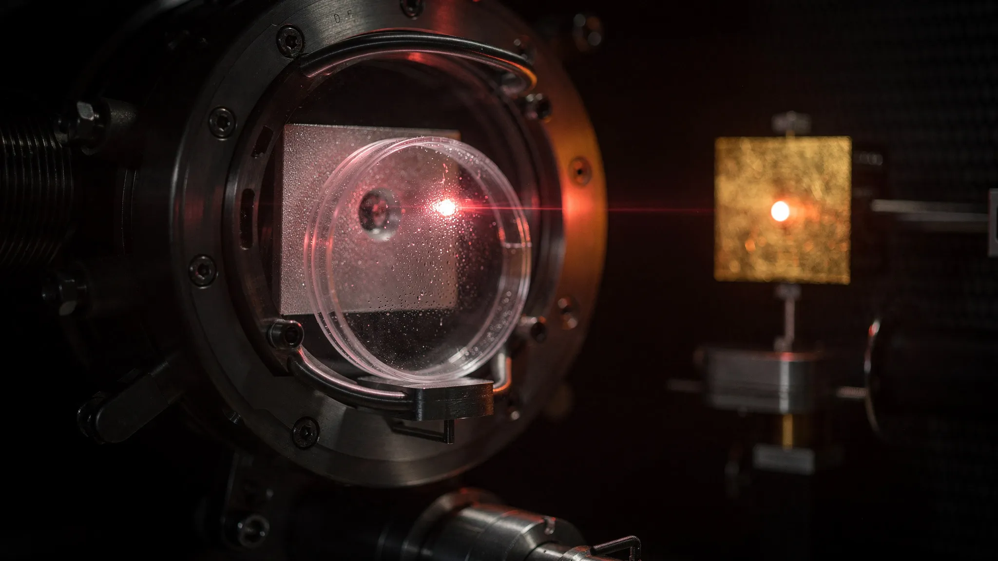
At the TARANIS laser facility in Belfast, pulses of 10.6 joules lasting 0.8 picoseconds strike a thin gold foil, producing a burst of electrons with energies of several million electronvolts. The beam spreads widely, so the dose reaching the cells is set simply by their distance from the source, between 33 and 75 mm. Each burst has stretched to about 12.5 to 23 picoseconds by the time it arrives, giving mean dose rates of 3 × 10¹⁰ to 5 × 10¹¹ grays per second, from a single pulse of up to about 8 grays. Film behind each sample measured the dose of every shot.
The cells were grown as thin single layers: two healthy human lines (RPE-1 and AG01522-D) and two cancer lines (DU145 and MCF7). The reference was an ordinary X-ray machine delivering 2.4 grays per minute. Twelve days later, the team counted how many cells had survived well enough to grow into colonies.
More survivors above 3 grays
Up to about 2 to 3 grays, the two treatments killed cells equally well: their relative biological effectiveness — the X-ray dose divided by the pulse dose that leaves the same fraction of cells alive — is 1.0 ± 0.1 at 50% survival. At higher doses, every cell line survived better after the picosecond pulse. To kill 90% of the cells, the pulse needed about 1 / 0.84 ≈ 1.2 times the X-ray dose: an effectiveness of 0.84 ± 0.05, comparable to FLASH-like sparing reported elsewhere. Two more cancer lines, HeLa S3 and H460, tested up to 2 and 3 grays, showed no difference either, consistent with the other lines.
Survival curves are usually described by two numbers: α, for damage that kills in one hit, and β, for damage that kills when sublethal hits accumulate — the term that grows with the square of the dose. The sparing came from β, roughly halved, while α stayed about the same. For the healthy RPE-1 cells, plugging the paper’s rounded fitted values into the survival formula, exp(−(αD + βD²)) for a dose D, gives about 8% survival at 6 grays after the pulse, against about 4% with X-rays.
Markers of DNA double-strand breaks (cuts through both strands of the DNA) showed far fewer damage sites half an hour to an hour after the pulse, but as many as with X-rays 24 hours later. The authors read this as similar repair, with no change in the hardest-to-repair damage — consistent with an unchanged α.
Healthy or cancerous, the same response
The most intriguing result is what did not vary. Healthy and cancer cells behaved alike, which suggests a mechanism rooted in early physics and chemistry rather than in the biology of a given cell type. Oxygen cannot move or be consumed by the cell in a few picoseconds. But the pulse overlaps the first instants of water being split by radiation, and the authors suggest that the reactive species produced could briefly use up oxygen over nanometre distances, so that fewer DNA lesions are made permanent.
The comparison with earlier work makes the puzzle sharper. A single femtosecond pulse, even shorter, also lowered β, but raised α, so overall it killed more cells, not fewer. Something seems to change abruptly between femtoseconds and picoseconds, and its cause is, in the authors’ words, currently unknown.
These are cells in dishes, irradiated in a single campaign, not patients. What the study offers is a new kind of laboratory: radiation delivered on the timescale of the earliest chemistry, against which the simulation codes of radiobiology, whose predictions vary widely today, can finally be tested.