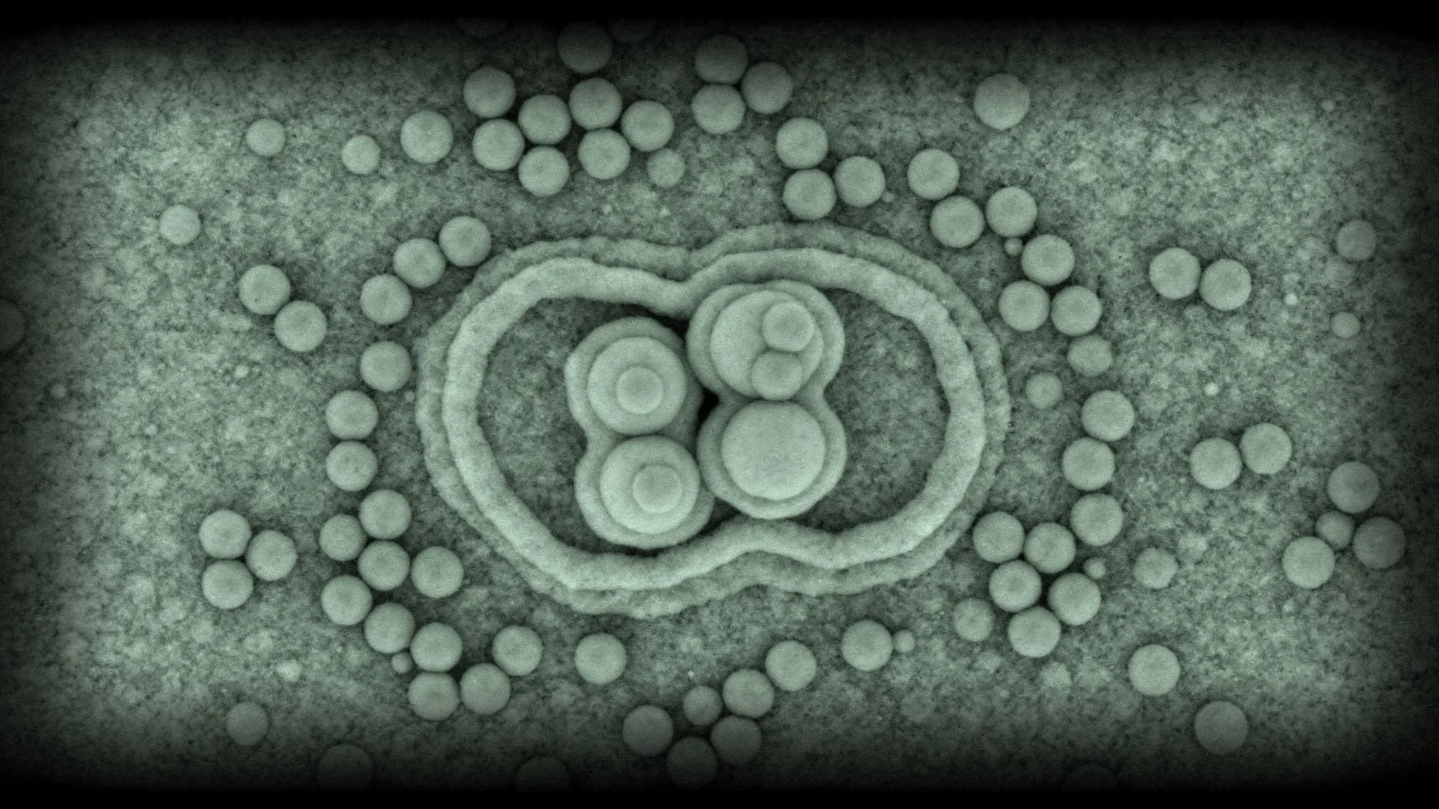
A VIRUS GETS A SECOND COAT, BUILT BY ELECTRIC ATTRACTION
Many simple viruses with an RNA genome build themselves. Infected cells churn out copies of a coat protein, and these copies assemble around the viral RNA into a protective shell, the capsid. For the bacteriophage MS2, a virus that infects the bacterium E. coli, this even works in a test tube: 180 coat proteins assemble around MS2’s RNA into a shell about 30 nanometres wide, with the same structure as the real virus.
What drives the assembly? Most studies of MS2 focus on a strong, specific bond between one region of its RNA and the coat protein. But shells also form around RNA lacking that region. Another candidate is plain electric attraction between charged groups on the RNA and on the protein, tuned by the salt in the solution. In MS2 the two effects are hard to tell apart.
The virus as its own template
LaNell Williams, Amelia Paine, Kellianne Kornick, Rees Garmann and Vinothan Manoharan, at Harvard University, Princeton University and San Diego State University, found a way to separate them. Instead of RNA, they offered the coat protein a whole MS2 virus as a template. Its RNA is locked inside the capsid, so no specific RNA bond is possible, and its size does not change with salt.
They mixed the viruses with pairs of coat proteins (dimers) at concentrations from 1.25 to 20 micromolar, and looked at the result after 10 minutes with an electron microscope, gels, light scattering and infection tests.
Shells that grow on shells
- Below 5 micromolar, nothing happened. From that concentration up, extra protein layers formed around the viruses.
- At 5 micromolar — 100 coat-protein pairs per virus, in principle enough for a whole second shell — only a few viruses had a layer, and it was incomplete.
- At 15 micromolar and above, most viruses looked fully wrapped in the microscope images, many with a third layer or more, and some pairs of viruses shared a single outer shell. Yet the authors also report many particles with no layer or only a partial one at 15 micromolar, and some bare viruses even at 20.
- Coat proteins alone, at 15 micromolar, built nothing without viruses present: the virus acts as a seed.
Charges and salt
Measured in the same conditions, the coat-protein pairs carry a positive apparent charge and the viruses a negative one. Salt matters: clean extra layers formed only around 100 millimolar of sodium chloride. Without salt, the viruses clumped together; at 1,000 millimolar, the shells came out patchy.
Was the virus’s maturation protein — the protein it uses to latch onto bacteria — acting as the starting point? No: extra layers also formed around virus-like particles — shells assembled in the test tube from MS2 RNA and coat protein — that lack it. The new layers do, however, sometimes cover it up. The more coat protein was added, the less the coated viruses stuck to the bacteria’s F-pili, the structures this protein binds to, and the fewer bacteria they infected. Since some coated viruses still stuck, the layers seem to land at random spots on the shell.
A coat that stops halfway
The authors see the extra layers as growing from a seed: a first patch must form, which needs enough protein, then it spreads. The threshold, 5 micromolar, is higher than the roughly 2 micromolar found earlier for shells growing around bare RNA, which fits a template with no specific bonds to offer.
Why do so many second coats stop before closing? The authors suggest an elastic cost: a capsid is stiffer than RNA, and an outer layer would need a different curvature than the shell it rests on. They conclude that electric attraction alone can drive shell assembly, even in a virus known for its specific RNA–protein bonds. Layered particles might one day help design sturdier virus-like particles, for example to carry drugs — but that remains a possibility, tested here only in a test tube.