A DECADES-OLD DEBATE ON BIOTECH'S STRONGEST BOND, SETTLED BY A PIPETTE
Streptavidin is a protein that grips a molecule called biotin with extraordinary force. Their dissociation constant — the biotin concentration at which half the binding sites are occupied; the lower it is, the tighter the grip — is around 4 × 10⁻¹⁴ molar, often cited as one of the strongest non-covalent interactions in biology. Combined with a very slow release and high specificity, this makes the pair a workhorse of nanobiotechnology, from immunoassays to immobilising molecules on surfaces.
Streptavidin is made of four identical subunits, with four binding sites for biotin. That raises an old question: are the sites cooperative? Does a biotin already in place make the next one bind more easily — or less? Over the decades, studies have reported positive cooperativity, negative cooperativity, and none. A frequently cited 1995 study found none, but only after letting the mixture reach equilibrium for two days, far longer than most modern applications wait.
The trouble with labels
Fluorescence microscopy can follow single molecules, so it seems ideal for watching biotin bind. But it usually requires attaching fluorescent dyes. On streptavidin, dyes sit nanometres apart and dim each other, which blurs the count of binding events, and dye coupling has been shown to impair biotin binding itself.
Malavika Kayyil Veedu, Jérôme Wenger and colleagues at the Fresnel Institute in Marseille (Aix-Marseille University, CNRS, Centrale Med) avoided labels entirely. They lit the protein with ultraviolet light at 295 nanometres and recorded the faint glow of its own tryptophan residues, building blocks of the protein.
Counting full proteins
The protocol counts only streptavidins carrying all four biotins. The team mixed 200 nanomolar streptavidin with increasing amounts of biotin, then added silica beads coated with biotin: any protein with a free slot sticks to them and is spun away. What remains in solution — the fully loaded proteins — is measured by its ultraviolet glow.
The shape of the resulting curve tells the story. If the sites are independent, the fraction of fully loaded proteins rises gradually as biotin is added. With positive cooperativity, biotin piles onto a few proteins first and the curve rises early and steeply; with negative cooperativity, full loading is delayed. Simulations, whose complete code is given in the paper, trace each case.
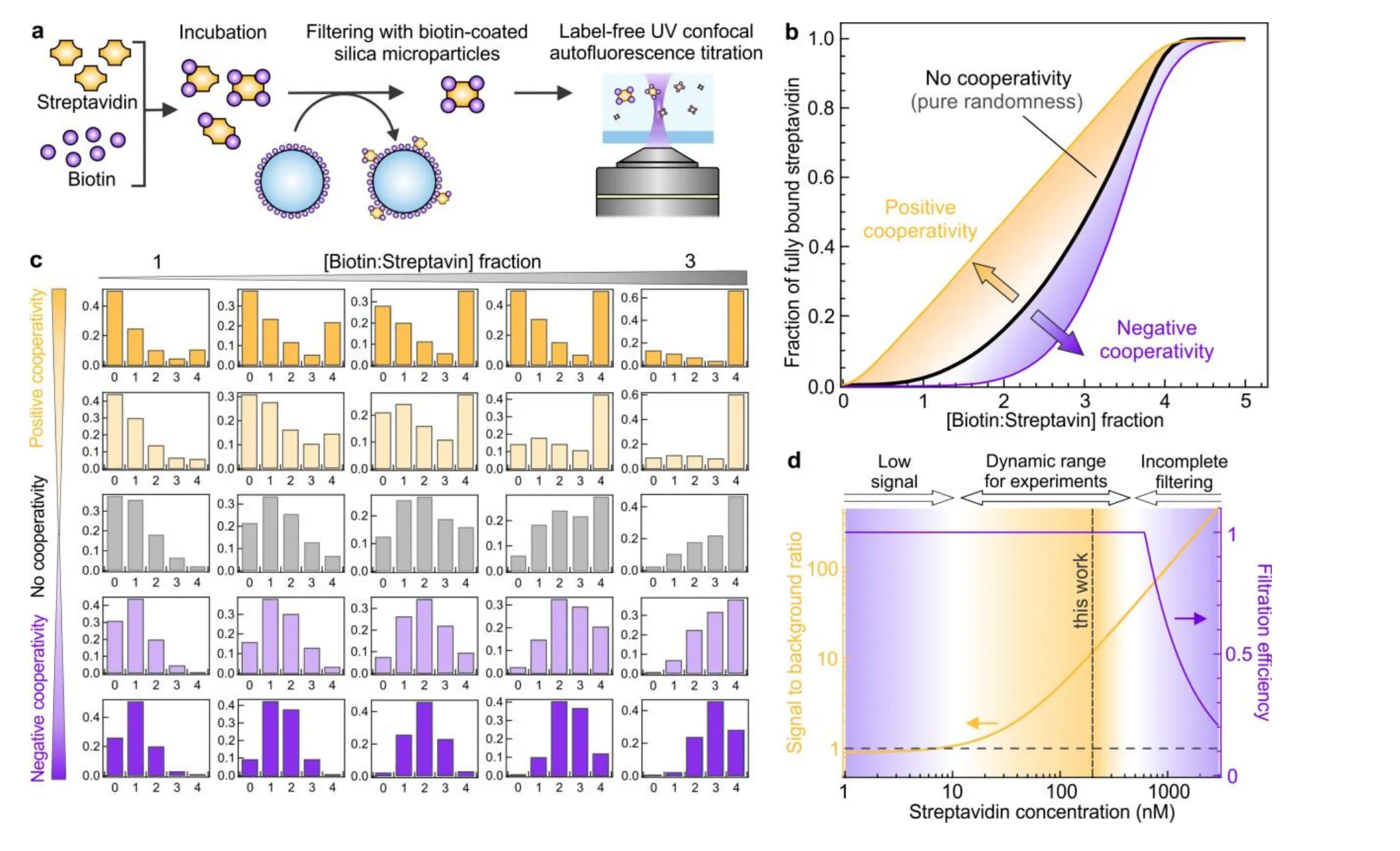
(a) The protocol: incubation, removal of incompletely loaded proteins on biotin-coated beads, label-free ultraviolet measurement. (b) Simulated fraction of fully loaded streptavidin for no, positive and negative cooperativity. (c) Simulated occupancy of the four sites. (d) The working range. — Figure 1, Kayyil Veedu et al. (2026), arXiv:2610.41363.
One experiment, two answers
The team ran the same experiment twice, changing only the concentration of the biotin solution they pipetted in. With dilute biotin (1.25 or 2.5 micromolar, added in volumes always above 15 microlitres), the data follow the curve for independent sites. With concentrated biotin (25 micromolar, added in drops of a few microlitres), the data fall squarely in the zone of positive cooperativity.
The protein cannot be both. The dilute data fit a model with no cooperativity, once a little imperfect filtering is allowed for. So what happened with the concentrated drops?
Faster than the stir
Biotin binds streptavidin very fast: at micromolar concentrations, within a few tens of milliseconds — much faster than a solution can be mixed. A small drop of concentrated biotin therefore reacts with the proteins around it before it has spread. Locally, there is far more biotin per protein than the average, so a subset of proteins fills up completely. On the curve, that looks exactly like cooperativity.
The team modelled this simply: the reaction happens inside a sphere of radius 1.5 R + 1 millimetre, where R is the radius of the added drop. The factor 1.5 comes from filming drops of black ink spreading in water; the extra millimetre was fitted to the data. With no cooperativity at all, this model reproduces the “cooperative” points.
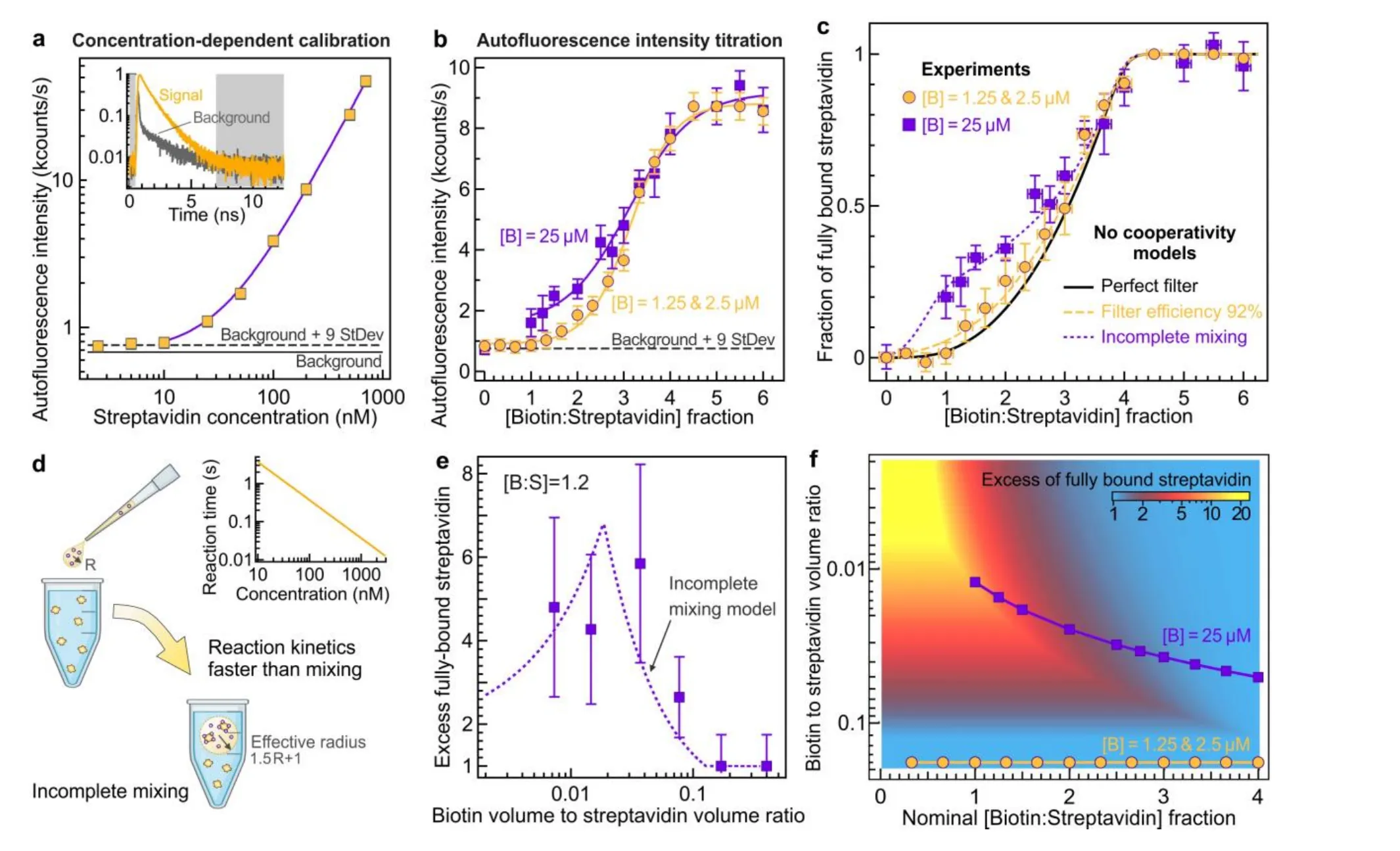
(c) Dilute biotin (orange) follows the curve without cooperativity; concentrated biotin (purple) seems cooperative, and is matched by the incomplete-mixing model. (d) The model. (e) Excess of fully loaded proteins as the added volume shrinks. (f) Map of conditions prone to the artefact. — Figure 2, Kayyil Veedu et al. (2026), arXiv:2610.41363.
A map for careful pipetting
To test the idea directly, the team fixed the ratio at 1.2 biotins per streptavidin and varied only the volume of the biotin added. As the drop shrank, the excess of fully loaded proteins, compared with a well-mixed reference, grew, peaking when the biotin volume was 2% of the protein volume — where, inside the reaction zone, the local ratio reaches about 4 biotins per protein, enough to fill every slot.
From the validated model, they drew a map. Adding biotin in a volume above 15% of the protein solution keeps the mixture clean. Small volumes at low ratios can inflate the number of fully loaded proteins more than fourfold.
What the protein really does
Biotin binding is known to make streptavidin more compact and more stable. The team concludes that these changes do not translate into cooperative binding under the conditions in which the pair is actually used. Their method cannot measure how many sites are filled on average, and they cannot rule out a very weak positive effect; but a result that changes with how the biotin is pipetted, and is fully explained without cooperativity, leaves cooperation little room. For labs that rely on this bond, the lesson is practical: when a reaction outruns the mixing, the size of the drop can write the result.