A FEW DNA DROPLETS, A MESH 25 TIMES STIFFER
Mix rigid particles into a soft material and it stiffens, in proportion to the room they take up. For small amounts the rule carries Einstein’s name: the stiffness is multiplied by 1 + 5ϕ/2, where ϕ is the fraction of the volume the particles fill. With a tenth of the volume, that is a factor of 1.25 — at most 25% stiffer, even for perfectly rigid particles.
Sam Wilken, at Johannes Gutenberg University Mainz, Omar Saleh, at the University of California, Santa Barbara, and colleagues used liquid droplets instead. Filling less than a tenth of the volume, they made a material about 25 times stiffer — reversibly.
Filaments from muscle, stars made of DNA
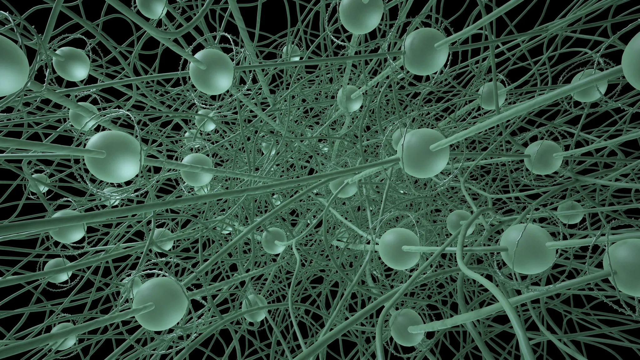
The matrix is a tangle of actin, purified from rabbit muscle powder. Its units join into filaments 10 to 30 micrometres long and only 7 nanometres thick. They resist bending: their persistence length, a measure of that stiffness, is about 17 micrometres, close to their own length. Entangled, they form a soft mesh.
The droplets are made of DNA nanostars, four-armed particles about 10 nanometres across. Their arms end in short “sticky” strands that bind to one another. Below a melting temperature — 27 to 31 °C depending on the mix — the stars gather into droplets of a dense DNA liquid, which dissolve again above it. This is liquid–liquid phase separation, and such biomolecular droplets are called condensates. Actin and DNA do not bind to each other.
Droplets that grow around the filaments
The team let the actin assemble at 37 °C, then cooled the sample. Without actin, droplets merged into large drops that sank. With actin, they stayed a few micrometres across, suspended throughout. Two-colour imaging held a surprise: the actin signal stayed uniform even inside them. The droplets had formed around the filaments instead of pushing them aside, so each one is threaded by actin — for more than 14 hours; three days later, the actin had mostly left.
The mesh also sets the droplets’ size. Droplets grow by jiggling about and merging; the mesh holds each one as if on a spring, so merging stops once no neighbour is within reach. The stiffer the mesh, the smaller the droplets, as the authors’ model predicts — and their sizes are more uniform than without actin.
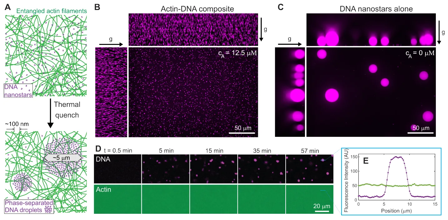
A: the actin mesh (green) and DNA nanostars (purple) before and after cooling. B: with actin, small droplets stay suspended throughout the sample. C: without actin, they grow large and sink (g marks gravity). D–E: the actin signal stays uniform, even across a droplet. — Figure 1, Wilken et al. (2026), arXiv:2610.38504.
Measuring stiffness by twisting
Stiffness was measured in a rheometer, which twists a thin layer of sample gently back and forth between two glass plates. Two numbers come out: the elastic modulus G′, the part of the resistance that springs back like a solid, and the loss modulus G″, the part lost as in a liquid. Both are in pascals, the unit of pressure — here, the stress needed to shear the sample per unit of deformation.
Twenty-five times, not twenty-five per cent
The actin mesh alone, at 20 µM (micromolar, a measure of concentration), had an elastic modulus of about 1 pascal when warm, twice that when cold. Adding 38 µM of nanostars, dissolved above the melting point, doubled it. Then, as droplets formed below about 31 °C, the modulus jumped and kept rising: on the team’s plot, it reaches roughly 100 pascals at 10 °C.
Divided by the modulus of the same mix without droplets, this gives a stiffening factor of about 25 at the lowest temperatures, with droplets filling only about 9% of the volume. At that fraction, Einstein’s rule allows a factor of 1.225 at most. And cycling the temperature across the melting point switches the material between soft and stiff, again and again, in about 10 minutes — the time droplets take to reach their final size.
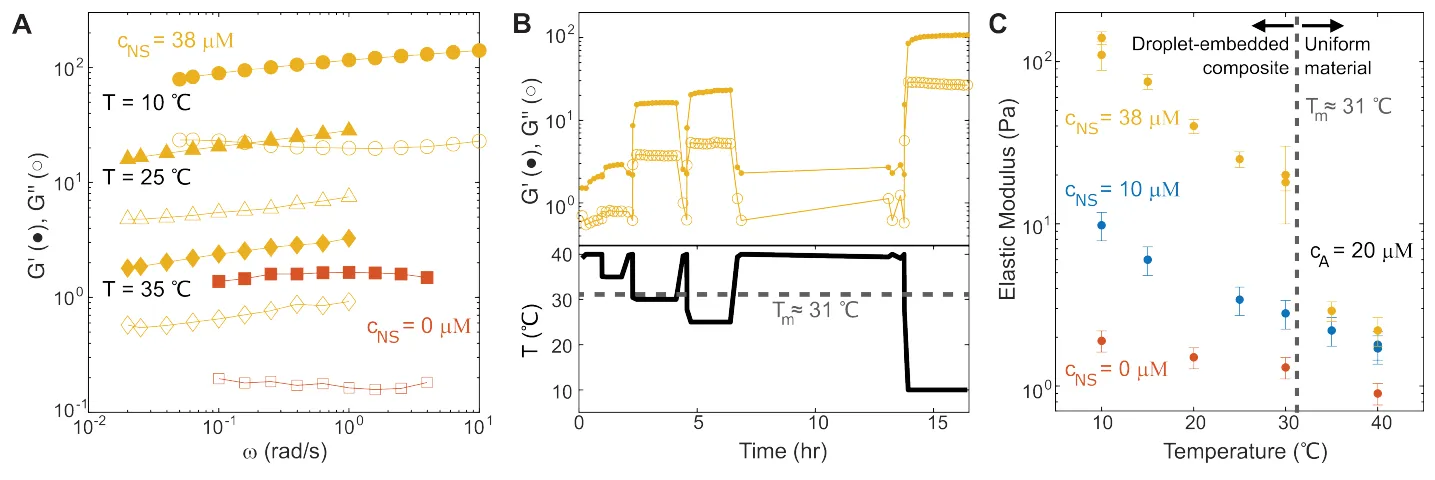
A: elastic (G′, filled) and loss (G″, open) moduli versus frequency, for actin alone (red) and with nanostars at three temperatures (yellow). B: as the temperature (bottom) is cycled across the melting point, the stiffness (top) switches back and forth. C: elastic modulus versus temperature, without nanostars and with 10 or 38 µM. — Figure 3, Wilken et al. (2026), arXiv:2610.38504.
Gripped filaments, dragged neighbours
Two usual explanations fail: the DNA liquid’s surface tension, the pull of the droplets’ surface, is far too weak, and nanostars gluing filaments together would make the material stiffen further under larger deformations — it did the opposite.
Inside a droplet, actin filaments are squeezed into a “tube” about 10 nanometres wide — barely more than their own 7 — against about 140 nanometres in actin alone, where each filament wriggles inside the gap its neighbours leave it. That tight grip turns each droplet and its filaments into a rigid lump. Still not enough: at 9% of the volume, even perfectly rigid lumps stay within Einstein’s limit.
The key is shear. Shearing a material both stretches it and turns it; a rigid lump cannot stretch, so it only turns. The filaments gripped inside turn with it, and their outer parts sweep through the surrounding mesh, dragging it along; a droplet 2 micrometres in radius holds about 500 of them. The authors assume the dragged zone extends one droplet radius beyond the surface. That doubles each lump’s effective radius and multiplies its effective volume by 8.
With 10 µM of nanostars, droplets fill 2.5% of the volume, so the enlarged lumps fill 20%. They stay separate; standard models then predict a stiffening factor of 1.8, close to the 2.2 ± 0.3 measured. With 38 µM, 9% becomes about 70%, far above the threshold — about 30% — at which such overlapping shells link into a continuous web, joined by shared filaments, that holds the material rigid. The stiffening stays finite: the web is no stiffer than the confined actin inside each droplet.
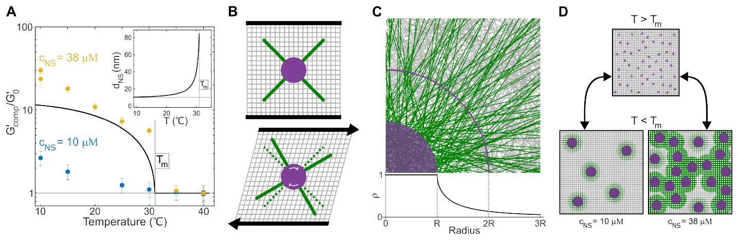
A: stiffening factor versus temperature; the line is the authors’ confinement prediction. B: under shear, a droplet (purple) rotates and drags the filaments threading it (green). C: near a droplet, most filaments are ones that cross it; the arc marks twice its radius. D: below the melting point, droplets with their dragged halo stay apart at 10 µM and join into a web at 38 µM. — Figure 4, Wilken et al. (2026), arXiv:2610.38504.
One droplet at a time
The picture rests on an assumption — a dragged zone exactly one radius thick — and treats each droplet alone. Where zones overlap, filaments shared by two droplets cannot turn with both, a stress the model leaves out. Whether the droplet size set by the mesh matters for the stiffening, or only the volume fraction, is unclear. And the curves come from two samples per nanostar concentration, one for actin alone.
The authors call the coupling “reciprocal, not feedback”: two one-way influences pointing in opposite directions. In biology, where condensates and filament networks are both widespread, forming droplets could change mechanics on demand, they suggest. For now, in this mix, the mesh decides how big the droplets grow, and the droplets decide how hard the mesh pushes back.