
HEMOGLOBIN MAY BE LESS COOPERATIVE THAN TEXTBOOKS SAY
Hemoglobin, the protein that carries oxygen in our blood, is also the textbook example of cooperativity: once it has bound one oxygen molecule, it binds the next ones more easily, up to four. This is what lets it load up fully in the lungs and unload in the tissues. Two classic models explain it — the one L. Pauling proposed in 1935, in which the four binding sites help one another, and the Monod-Wyman-Changeux (MWC) model of 1965, in which the whole molecule switches between two shapes that hold oxygen more or less tightly.
Both are usually worked out in the same setting: a single hemoglobin molecule immersed in a reservoir of free oxygen molecules, described by a pressure. Physicists call this the “grand canonical ensemble”. The saturation curve — how much oxygen hemoglobin holds at each pressure — follows from it, and cooperativity is read off that curve.
A reservoir that isn’t there
A. E. Allahverdyan and colleagues, at the Alikhanyan National Laboratory and Yerevan State University in Armenia, point out that inside a red blood cell, that reservoir does not exist. Free oxygen dissolved in the cell is at least 100 times less concentrated than oxygen bound to hemoglobin — which is the very reason hemoglobin exists. There are about 30 hemoglobin molecules for every free oxygen molecule. Even in arterial blood, most hemoglobin molecules have no free oxygen anywhere near them: each one sees only the four molecules it holds.
So the team describes the situation differently: one hemoglobin and a handful of oxygen molecules — five at most — in a fixed volume, the “canonical ensemble”. That volume is set by how tightly hemoglobin is packed in the cell: about four times the molecule’s own volume, a cube 6.93 nanometres on a side. In this picture, oxygen moves in or out according to differences in free energy, a measure of which arrangement is statistically favoured.
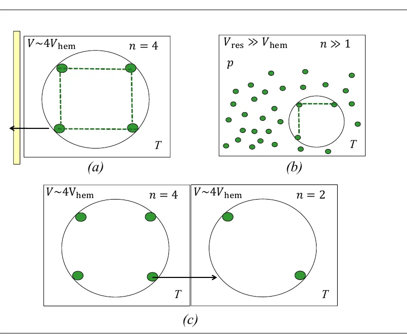
(a) The new picture: one hemoglobin (circle) with its four oxygen molecules in a volume about four times its own. (b) The usual picture: a hemoglobin in a reservoir of many free oxygen molecules. (c) Two neighbouring hemoglobins can exchange oxygen. — Figure 1, Allahverdyan et al. (2026), arXiv:2611.00824.
Weaker cooperativity
The team fitted both classic models to standard data on how hemoglobin releases oxygen as the oxygen pressure drops — the first molecule leaving between 80 and 40 torr (a unit of pressure; in the lungs, oxygen sits at about 100 torr), the last between 20 and 7.5 torr.
- In Pauling’s model, the cooperativity factor — how much a bound oxygen boosts binding at a neighbouring site; 1 means no boost — comes out at 1.88 in the new framework, against 6.32 with the usual one, fitted to the same data. Pauling’s own fit, with older data, gave 12.
- In the MWC model, the ratio between the oxygen affinities of the two shapes — a measure of how far the molecule departs from simple, independent binding — comes out at about 4.7, against 95.4 in the usual framework.
Some cooperativity is still needed. Without it, hemoglobin could load up in the lungs but would only serve tissues at rest.
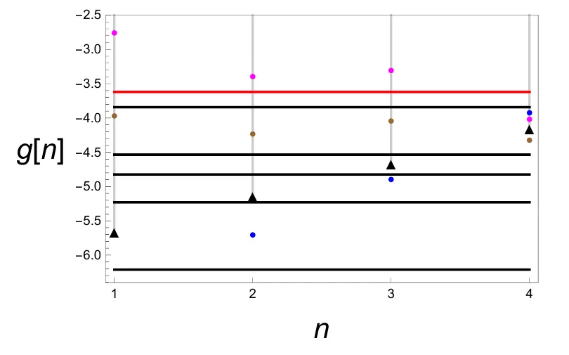
Free-energy levels of Pauling’s model for one to four bound oxygen molecules. The horizontal lines mark the physiological windows; only the best fit (black triangles, cooperativity 1.88) falls in all of them. The other colours show stronger cooperativity (magenta, brown) and none (blue). — Figure 2, Allahverdyan et al. (2026), arXiv:2611.00824.
When cooperation turns into a cascade
In the new framework, too much cooperativity becomes dangerous. With a factor of 4.83, the calculation produces a cascade: as soon as the first oxygen molecule leaves, the other three are released automatically — a scenario the authors call unacceptable, because it would lead to oxygen poisoning. At 7.83, hemoglobin could no longer load up in the lungs at all. The values of the usual framework, imported into the new one, likewise lead to instabilities. The usual framework, by contrast, sets no such limits.
The new picture also changes the character of the molecule. In the usual description, the number of bound oxygen molecules fluctuates about as much as its average value. In the canonical one, it barely fluctuates: hemoglobin behaves almost like a machine, taking and releasing oxygen one step at a time, and a full molecule attracts no more.
Oxygen that hops
The authors draw one more consequence: since neighbouring hemoglobins can trade oxygen according to their free energies, oxygen might travel inside the red blood cell partly by hopping from one hemoglobin to the next. Testing that idea will take dynamic models. The study is theoretical, and its physiological thresholds are, in the authors’ words, “somewhat conventional” — but it questions a cornerstone of how biology textbooks describe the oxygen in our blood.